Hospital Hours: Monday-Friday: 8am-6pm | Saturday: 8am-4pm
(519) 896-0532


Most lumps people call sebaceous cysts in dogs are harmless, and most aren't actually sebaceous. They come from hair follicles, not oil glands, and they fill with a thick, cheesy material. A lump's look and feel can't tell you what it is, though, so I'd have any new one checked with a needle sample.
Key Takeaways
Book a visit soon if a lump is growing quickly, changing color, bleeding or painful, if your dog keeps licking or chewing at it, if one has burst, or if several new lumps appear. Call (519) 896-0532 or book online. Outside our hours, go to the nearest emergency hospital.
A sebaceous cyst is a closed sac under the skin filled with a thick, cheesy material. The proper name is usually follicular cyst, and you'll also hear infundibular or epidermal cyst. Merck describes most keratinized skin cysts in dogs as malformations of the hair follicle.1
The contents are keratin, the protein that makes up hair and the outer skin. It's gray, brown or yellowish and crumbly, like soft cheese.1 Cysts are usually single, freely movable under the skin and slightly compressible. They run from a couple of millimeters to more than 5 cm.1
True sebaceous gland growths exist too, but they're a separate group of tumors that are common in older dogs and often multiple.2 A needle sample or biopsy is how we tell them apart.
The sac forms when a hair follicle develops abnormally or gets plugged, and keratin builds up inside with nowhere to go.1 Several things make that more likely.
Most appear on the head, neck, trunk and upper limbs. They can show up in the ear canal and around the rear end too.
Cysts are usually single, but some dogs grow several. In young dogs, a cluster on top of the head may be congenital. A lump between the toes is a different problem, and we cover those in our post on interdigital cysts.
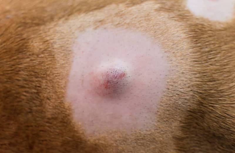
A cyst feels like a well-defined bump, firm to soft, sitting in or just under the skin. Many have a small pore or dark plug at the center. Some look slightly bluish.
A cyst that bursts releases a thick yellow-brown or gray material. Inflamed ones can be sore or itchy, and the skin around them may turn red and swollen. If bacteria get in, you can end up with a secondary skin infection, which we explain in our post on pyoderma.


You can't tell by looking or feeling. A lipoma, an abscess and a mast cell tumor can all pass for a cyst, and Merck says identifying skin tumors should be left to a veterinarian.2
| Lump | How it often feels | Other clues | What I'd do |
|---|---|---|---|
| Follicular ("sebaceous") cyst | Round, firm to soft, movable | May have a central pore. Releases cheesy material if it bursts.1 | Aspirate. Then watch it or remove it. |
| Lipoma | Soft, movable, often on the trunk or upper legs3 | Benign fat tumor, most common in older, overweight dogs2 | Aspirate. Excision is curative if you want it gone.3 |
| Abscess | Warm and painful | Often follows a bite or wound. May drain pus. | Needs treatment. Book a visit soon. |
| Mast cell tumor | Varies. Can mimic a cyst or a lipoma. | The most common malignant skin tumor in dogs2 | Aspirate before anything else. |
Diagnosis starts with an exam and, in most cases, a needle sample. I would suggest the needle sample for any lump we haven't identified before.
Cytology is reliable but not perfect. In a veterinary pathology study comparing aspirates with final lab results, the aspirate matched the diagnosis in 90.9% of cases. When it called a lump "not a tumor," it was right 68.7% of the time.5 That's why I'd suggest removing any lump that keeps changing or that the aspirate can't explain, and sending it to a lab.
Once a lump is confirmed as a cyst, you have two reasonable options: remove it or watch it. Excision is the treatment of choice,1 but cysts are benign, so watching is fine for a small one that isn't bothering your dog.
I would recommend removal if a cyst is growing, keeps rupturing, gets infected, sits where it rubs, or if we can't be sure what it is. Removal isn't always practical for a dog with dozens of them. If surgery makes sense, you can read about surgery at Kingsdale.
If you're watching one, measure it and take a photo next to a coin once a month. A change in size, color or feel is a reason to book a recheck.
Don't squeeze it. Squeezing can push keratin into the surrounding tissue, which sets off a heavy foreign body reaction and often turns a quiet lump into a sore one.1,4
A true cyst is a benign sac and doesn't turn into cancer. The risk is a different lump that looks like a cyst. Lipomas, abscesses and mast cell tumors can all feel similar, and mast cell tumors are the most common malignant skin tumor in dogs. A fine needle aspirate tells them apart, so I would suggest having any new or changing lump checked rather than assuming it's a cyst.
Yes. Boxers, Shih Tzus, Miniature Schnauzers, Old English Sheepdogs, Rottweilers, Doberman Pinschers and Bullmastiffs are reported as predisposed. Any dog can get one, and some dogs grow several.
No. Squeezing can push the cyst's keratin into the surrounding tissue, which triggers a heavy inflammatory reaction and often an infection. If a cyst has burst on its own, stop your dog from licking it and call us so we can have a look.
No. Removal is the treatment of choice, but once a lump is confirmed as a cyst it's reasonable to watch a small one that isn't bothering your dog. I would recommend removal if it's growing, keeps rupturing, gets infected, sits where it rubs, or if we can't be sure what it is.
Sources


