


Calcinosis cutis, or cutaneous calcification, is a condition that can affect dogs and cause lesions or bumps to form on the skin. These bumps are usually red, white, or yellow and can be uncomfortable or distressing to your pet. Diagnosing and treating calcinosis cutis in dogs will depend on the severity of the condition, but there are steps you can take as a veterinarian to manage this issue effectively. In this article, we’ll discuss calcinosis cutis, common symptoms in dogs, diagnosis, and treatment options for managing it successfully.
Calcinosis cutis in dogs is a skin disorder where calcium builds up in the dermis and beneath the skin, along with collagen and elastin fibers. This build-up of calcium can cause very firm bumps to form on the skin. The affected skin of dogs with calcinosis cutis contains clusters of mineral deposits that can range from localized to widespread and from multifocal to coalescing.
Calcinosis cutis is mainly caused by the long-term use of corticosteroids such as prednisone in dogs, as well as conditions such as Cushing's disease and kidney failure. It can also be associated with certain infectious diseases, like leptospirosis and systemic blastomycosis, but it can be difficult to determine the cause in those cases. Certain breeds, including the German shepherd, English bulldog, Labrador retriever, Boston terrier, boxer, and rottweiler are predisposed to developing calcinosis cutis.
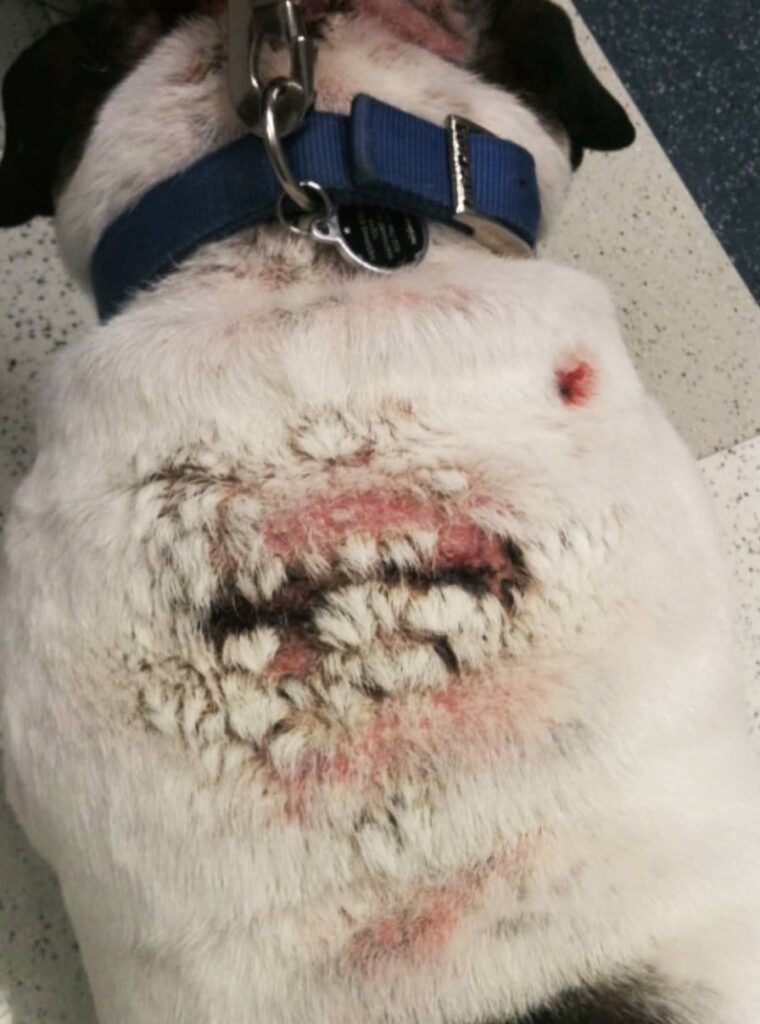
Calcinosis cutis in dogs may be characterized by:
Calcinosis cutis in dogs commonly affects the back, groin, and shoulder regions. Dogs may experience intense itchiness, and their lesions may be complicated by secondary bacterial and/or fungal infections.
Your veterinarian will perform a thorough physical examination of the affected area and may recommend taking a sample of the affected skin for laboratory testing. This may include a cytology (microscopic examination of cells) and/or a biopsy (removal and examination of a small piece of tissue) to confirm the diagnosis.
Your veterinarian should conduct skin cytology to identify potential bacterial or yeast infections that could be contributing to the clinical signs. These secondary infections may require medical treatment, so they must be addressed appropriately.
A presumptive diagnosis of calcinosis cutis can usually be made based on the dog's history and an examination, but to truly confirm the diagnosis of calcinosis cutis in dogs, it requires a biopsy. Fortunately, this procedure can be done with sedation rather than requiring a full general anesthetic. The biopsy sample will be sent to a veterinary pathologist for examination under a microscope.
To ensure an optimal outcome, it is important to accurately identify and manage any underlying causes of calcinosis cutis to prevent further progression.
The most studied treatment for calcinosis cutis in dogs is dimethyl sulfoxide (DMSO). Applying a thin layer of DMSO to affected lesions every 24 hours can be an effective way to break down calcium deposits and stop future crystallization.
Dog owners can use either liquid or gel-based products of DMSO, with gel-based products being simpler to apply. If your dog has large areas of skin affected by lesions, start by treating a small area, then gradually expand the treated area. Treating too much too quickly may have adverse effects, so it is important to progress gradually.
Using DMSO may result in the reabsorption of calcium into the bloodstream, which could lead to hypercalcemia, so make sure to periodically check calcium levels. Additionally, any secondary infections must be treated before applying DMSO. Treating associated bacterial or yeast infections can help reduce itching and discomfort while increasing the chances of lesion resolution.
Regular physical assessments are necessary to assess the progress of lesions, which can take weeks or even months to improve. Generally speaking, the prognosis for calcinosis cutis in dogs is good as long as a diagnosis is accurately made and the appropriate treatment is implemented.
Calcinosis cutis in dogs is a condition that can be diagnosed with physical examination, skin cytology, and biopsy. The most common cause of calcinosis cutis in dogs is a result of chronic corticosteroid use such as prednisone. Treatment involves the use of dimethyl sulfoxide (DMSO) to break down calcium deposits and stop further crystallization. To ensure an optimal outcome, it is important to accurately identify and manage any underlying causes of calcinosis cutis to prevent further progression. With proper diagnosis and treatment, calcinosis cutis has a good prognosis for dogs. If you have any questions about calcinosis cutis in dogs, please reach out to your veterinarian.


